CBCT Imaging
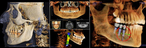
Cone-beam computed tomography systems (CBCT) are a variation of traditional computed tomography (CT) systems. The CBCT systems used by dental professionals rotate around the patient, capturing data using a cone-shaped X-ray beam. These data are used to reconstruct a three-dimensional (3D) image of the following regions of the patient’s anatomy: dental (teeth); oral and maxillofacial region (mouth, jaw, and neck); and ears, nose, and throat (“ENT”).
Uses :
Dental CBCT systems have been sold in the United States since the early 2000s and are increasingly used by radiologists and dental professionals for various clinical applications including dental implant planning, visualization of abnormal teeth, evaluation of the jaws and face, cleft palate assessment, diagnosis of dental caries (cavities), endodontic (root canal) diagnosis, and diagnosis of dental trauma.
Benefits:
X-ray imaging, including dental CBCT, provides a fast, non-invasive way of answering a number of clinical questions. Dental CBCT images provide three-dimensional (3-D) information, rather than the two-dimensional (2-D) information provided by a conventional X-ray image. This may help with the diagnosis, treatment planning and evaluation of certain conditions.